
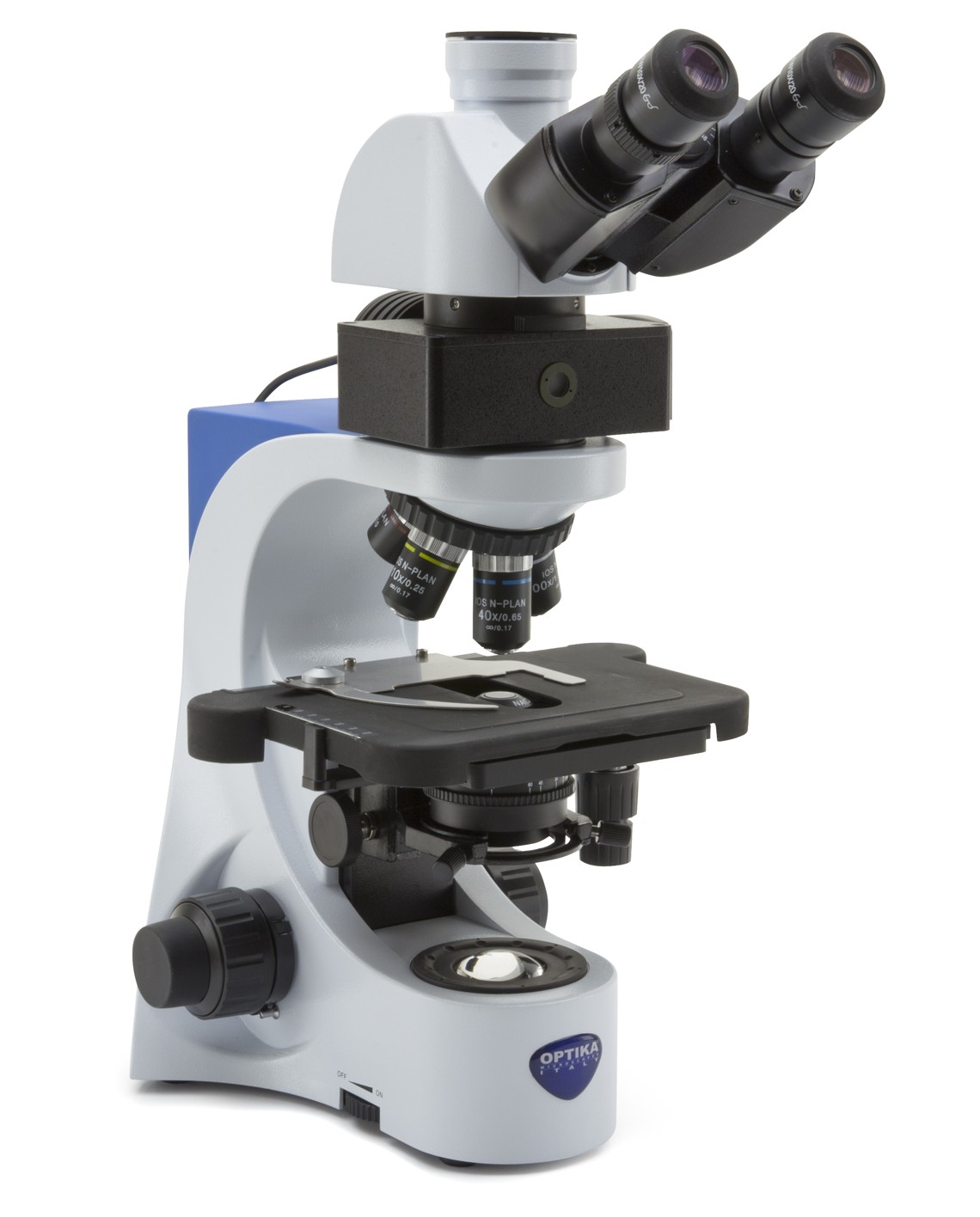


Optika B-383LD Trinocular LED Fluorescence Microscope, 1000x, IOS PLAN, B Filter Set, Multi-plug

Observation mode: Brightfield, LED fluorescence.
Epi-illumination and filter: High-power blue LED with brightness control. 3-position filter holder; blue included.
Head: Trinocular (fixed 50/50), 30° inclined, 360° rotating.
Interpupillary distance: Adjustable between 48 and 75 mm.
Dioptric adjustment: On the left eyepiece tube.
Eyepieces: WF10x/20 mm, high eye-point and secured by screw.
Nosepiece: Quintuple revolving nosepiece, rotation on ball bearings.
Objectives:
IOS N-PLAN 4x/0.10
IOS N-PLAN 10x/0.25
IOS N-PLAN 40x/0.65
IOS N-PLAN 100x/1.25 (Oil/Water)
All with anti-fungus treatment.
Specimen stage: Double layer rackless mechanical stage, 233×147 mm, 78×54 mm X-Y range.
Focusing: Coaxial coarse (adjustable tension) and fine focusing mechanism with limit stop to prevent the contact between objective and specimen.
Condenser: Abbe N.A. 1.25, with objective-coded iris diaphragm, focusable and centerable.
Transmitted illumination (Fixed Koehler type): X-LED3 with white 3.6 W LED (6,300K) with brightness control. Multi-plug 100-240Vac/6Vdc external power supply.
FLUO Series
Epi Fluorescence microscopes
A fluorescence microscope is an optical microscope that uses fluorescence and phosphorescence instead of, or in addition to, reflection and absorption to study properties of organic or inorganic substances. The “fluorescence microscope” refers to any microscope that uses fluorescence to generate an image. The Epi Fluorescence microscope is equipped with a fluorescence illuminator wich generates incident fluorescence light.highlight points of interest.
Principle
The specimen is illuminated with light of a specific wavelength (or wavelengths) which is absorbed by the fluorophores, causing them to emit light of longer wavelengths (i.e., of a different color than the absorbed light). The illumination light is separated from the much weaker emitted fluorescence through the use of a spectral emission filter. Typical components of a fluorescence microscope are a light source (HBO mercury-vapor lamps are common; more advanced forms are high-power LEDs), the excitation filter, the dichroic mirror, and the emission filter. The filters and the dichroic mirror are chosen to match the spectral excitation and emission characteristics of the fluorophore used to label the specimen. In this manner, the distribution of a single fluorophore (color) is imaged at a time. Multi-color images of several types of fluorophores must be composed by combining several single-color images. Most fluorescence microscopes in use are epifluorescence microscopes, where excitation of the fluorophore and detection of the fluorescence are done through the same light path (through the objective). These microscopes are widely used in biology and are the basis for more advanced microscope designs.
Epifluorescence microscopy
The majority of fluorescence microscopes, especially those used in the life sciences, are of the epifluorescence design. Light of the excitation wavelength illuminates the specimen through the objective lens. The fluorescence emitted by the specimen is focused to the detector by the same objective that is used for the excitation which for greater resolution will need objective lens with higher numerical aperture. Since most of the excitation light is transmitted through the specimen, only reflected excitatory light reaches the objective together with the emitted light and the epifluorescence method therefore gives a high signal-to-noise ratio. The dichroic beamsplitter acts as a wavelength specific filter, transmitting fluoresced light through to the eyepiece or detector, but reflecting any remaining excitation light back towards the source.
FLUORESCENCE
The fluorescence microscopy is the most demanding technique in biology and the biomedical sciences, as well as in materials science.
This method is capable to study organic and inorganic samples thanks to primary fluorescence (auto-fluorescence) or secondary (staining and labelling with fluorochromes).

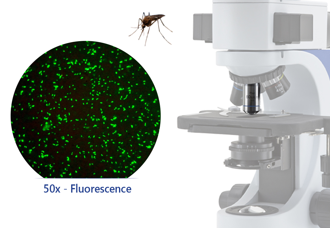
VALUABLE SUPPORT TO MALARIA & TBC DIAGNOSIS
The B-383LD1 LED fluorescent microscope is a cutting-edge solution designed to enable rapid diagnosis of malaria and TBC, by using acridine-orange staining technique. The optional no cover glass W-PLAN 50x/0.75 objective is required to ensure excellent results for stunning images.
The aim is to reduce the costs to a minimum and provide a cost-effective, intuitive but performing solution, purposely designed for these applications.
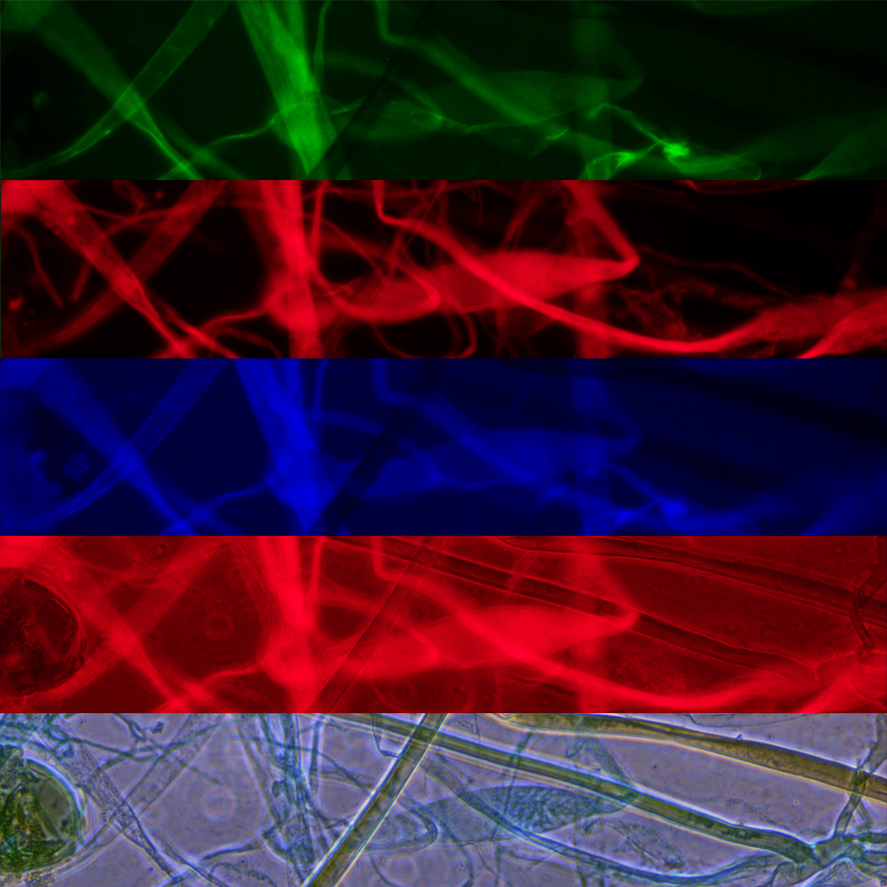
MULTIPLE OBSERVATION METHODS AVAILABLE
IM-5FLD means extreme flexibility.
In fact, brightfield, phase contrast, darkfield and fluorescence techniques can be used at any time, to highlight different aspects of your specimen.
Here you can see a Rhizopus sample, seen under (from top to bottom): Blue Excitation, Green Excitation, Ultraviolet Excitation, combination of Green Excitation and Phase Contrast, Phase Contrast
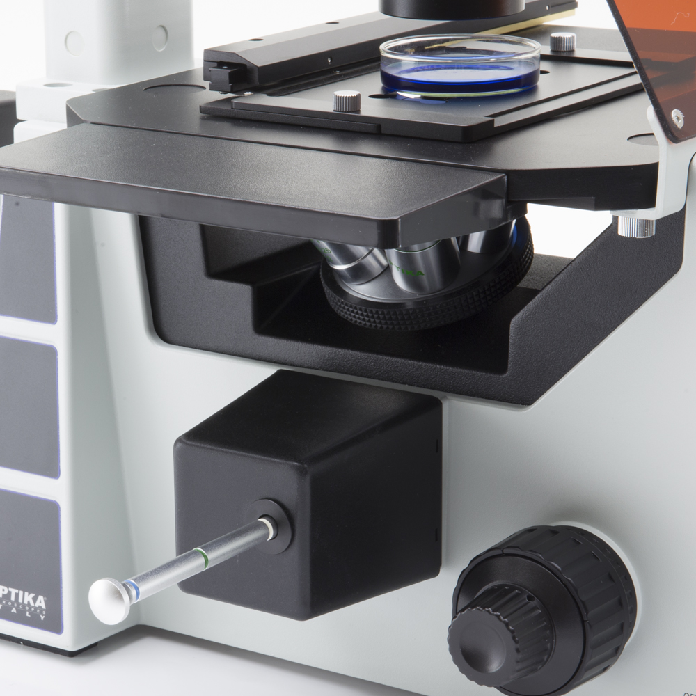
MOTORIZED LED SOURCE SELECTION
Using LED fluorescence has never been so easy!
With IM-5FLD, you will simply choose which filter is needed (among blue, green and UV) and then the microscope will select the most suitable lighting source for you, automatically.
In addition, an extra slot is available for the most demanding users, with the possibility to create a customized configuration.

